
Project: FMZ
Postmortem studies in schizophrenia reveal alterations in gene products that regulate the release and extracellular persistence of GABA. However, results of in vivo studies of schizophrenia measuring total tissue GABA with magnetic resonance spectroscopy (MRS) have been inconsistent. Neither the postmortem nor the MRS studies directly address the physiological properties of GABA neurotransmission. The goal of this line of research was to develop a mechanism for the in vivo measurement of GABA neurotransmission such that it could be utilized in the study of psychiatric disease, including schizophrenia.
In the first study, we measured the ability to increase GABA in eight healthy subjects by comparing the binding of [11C]flumazenil, a positron emission tomography (PET) radiotracer specific for the benzodiazepine (BDZ) site, at baseline and in the presence of an acute elevation in GABA levels through the blockade of the GABA membrane transporter (GAT1). Preclinical work suggests that increased GABA levels enhance the affinity of GABA-A receptors for BDZ ligands (termed 'GABA shift'). Theoretically, such an increase in the affinity of GABA-A receptors should be detected as an increase in the binding of a GABA-A BDZ-receptor site-specific PET radioligand. GAT1 blockade resulted in significant increases in mean [11C]flumazenil-binding potential (BP(ND)) over baseline in brain regions representing the major functional domains of the cerebral cortex: association cortex +15.2+/-20.2% (p=0.05), sensory cortex +13.5+/-15.5% (p=0.03) and limbic (medial temporal lobe, MTL) +16.4+/-20.2% (p=0.03). Moreover, the ability to increase GABA strongly predicted (r=0.85, p=0.015) the ability to entrain cortical networks, measured through EEG gamma synchrony during a cognitive control task in these same subjects.
Subsequently, this technique was utilized in a study of 17 off-medication patients with schizophrenia and 22 healthy comparison subjects. [11C]Flumazenil distribution volume (VT) was significantly increased across all cortical brain regions in the healthy comparison group but not in the schizophrenia group. This lack of effect was most prominent in the antipsychotic-naive schizophrenia group. In this subgroup, [11C]flumazenil ΔVT in the medial temporal lobe was correlated with positive symptoms, and baseline [(11)C]flumazenil VT in the medial temporal lobe was negatively correlated with visual learning. In the healthy comparison group but not the schizophrenia group, [11C]flumazenil ΔVT was positively associated with gamma-band oscillation power. This study demonstrates, for the first time, an in vivo impairment in GABA transmission in schizophrenia, most prominent in antipsychotic-naive individuals. The impairment in GABA transmission appears to be linked to clinical symptoms, disturbances in cortical oscillations, and cognition.
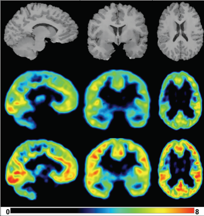
An acute increase in extracellular GABA levels within the cortex is detectable via increased specific binding of the radiotracer, [11C]flumazenil. Moreover, administration of tiagabine did not affect the plasma free fraction of [11C]flumazenil nor did it affect the free and non-specific binding of the radiotracer in the brain (measured as pons VT). Effects of tiagabine on [11C]flumazenil binding. MRI (top) and coregistered parametric BPND (unitless) maps measured under baseline (middle) and 30 minutes post-tiagabine (bottom) following [11C]flumazenil injection in a female volunteer. Parametric maps were obtained by derivation of BPND in each voxel with SRTM using the pons as the reference region. Increases in BPND were seen across all cortical regions.
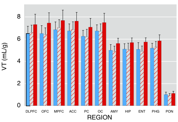
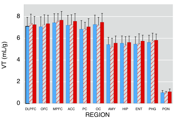
[11C]flumazenil regional distribution volumes (VT) at baseline (Top Panel) and post-tiagabine (Bottom Panel) for HC (blue bars, n = 22), DF SCH (red hatched bars, n = 9) and DN SCH (red bars, n = 8). Values are mean ± SD. ROIs include the dorsolateral prefrontal (DLPFC), orbital frontal (OFC), medial prefrontal (MPFC) and anterior cingulate (ACC) cortices; the Sensory Cortex, composed of the parietal (PC) and occipital (OC) cortices; and the limbic Medial Temporal Lobe (MTL) composed of the amygdala (AMY), hippocampus (HIP), entorhinal cortex (ENT) and parahippocampal gyrus (PHG).
No difference was observed when comparing [11C]flumazenil ΔVT between HC and SCH subjects (RM ANOVA F=1.09, df=1,35, p=0.30). However, separating the SCH group by prior treatment revealed that DN SCH subjects had significantly lower ΔVT compared with the healthy HC group (RM ANOVA F=5.93, df=1,26, p=0.02). This difference reached significance in the cortical regions DLPFC, OFC, MPFC and PC.
- In vivo measurement of GABA transmission in healthy subjects and schizophrenia patients.
Frankle WG, Cho RY, Prasad KM, Mason NS, Paris J, Himes ML, Walker C, Lewis DA, Narendran R.
Am J Psychiatry. 2015 Nov 1;172(11):1148-59. doi: 10.1176/appi.ajp.2015.14081031. Epub 2015 Jul 2.
PMID: 26133962 Free PMC article.
- [11C]flumazenil binding is increased in a dose-dependent manner with tiagabine-induced elevations in GABA levels.
Frankle WG, Cho RY, Mason NS, Chen CM, Himes M, Walker C, Lewis DA, Mathis CA, Narendran R.
PLoS One. 2012;7(2):e32443. doi: 10.1371/journal.pone.0032443. Epub 2012 Feb 27.
PMID: 22384252 Free PMC article.
- Human biodistribution and dosimetry of the PET radioligand [11C]flumazenil (FMZ).
Laymon CM, Narendran R, Mason NS, Carney JP, Lopresti BJ, Mathis CA, Mountz JM, Sashin D, Frankle WG.
Mol Imaging Biol. 2012 Feb;14(1):115-22. doi: 10.1007/s11307-011-0478-2.
PMID: 21365327
- Tiagabine increases [11C]flumazenil binding in cortical brain regions in healthy control subjects.
Frankle WG, Cho RY, Narendran R, Mason NS, Vora S, Litschge M, Price JC, Lewis DA, Mathis CA.
Neuropsychopharmacology. 2009 Feb;34(3):624-33. doi: 10.1038/npp.2008.104. Epub 2008 Jul 9.
PMID: 18615011 Free PMC article.